Image Gallery

PA NETs
Neutrophil extracellular traps of human neutrophils released in response to the bacterium Pseudomonas aeruginosa. Red: neutrophil DNA, green: bacteria (GFP-PAO1). Confocal microscopy performed by Balazs Rada

Human NETs
Neutrophil extracellular traps of human neutrophils stained for DNA (DAPI, blue), citrullinated histone H3 (red) and myeloperoxidase (MPO) – Performed by Dae-goon Yoo

Human NETs - 2
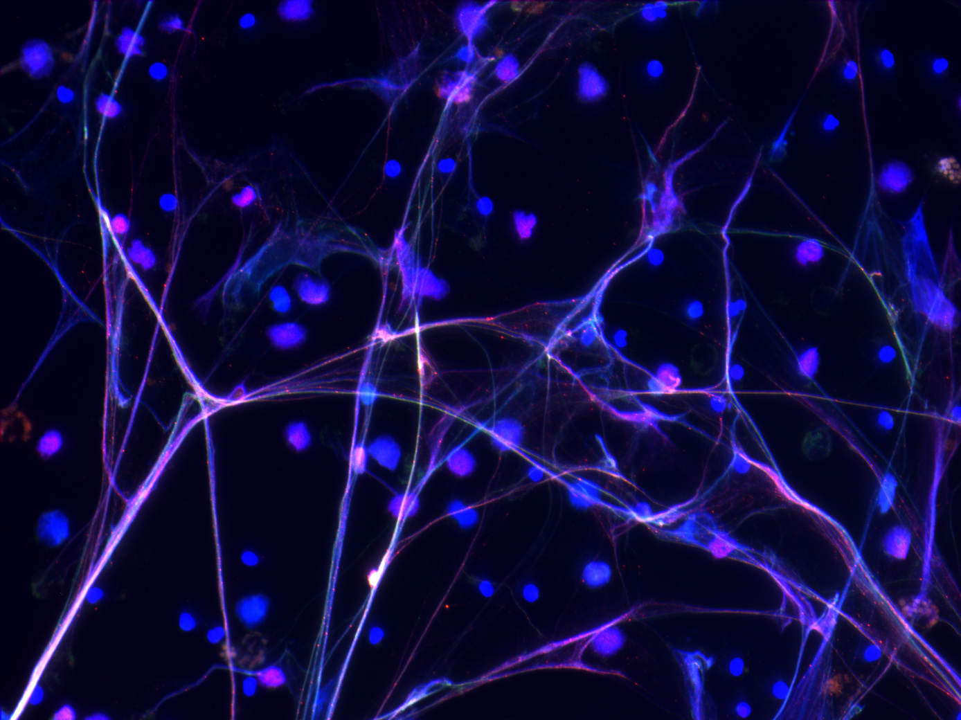
Neutrophil extracellular traps of human neutrophils stained for DNA (DAPI, blue) and neutrophil elastase (red) – Performed by Madison Floyd

Lactoperoxidase in mouse tracheaLactoperoxidase detection (brown) in murine airways by immunohistochemistry – Performed by Eszter Toth and Demba Sarr

Flu in mouse lung
Influenza virus in murine airways – detection of the viral nucleoprotein (brown) in airway epithelial cells and pulmonary macrophages by immunohistochemistry performed by the UGA Veterinary Diagnostic Laboratory. Influenza PR8 infection performed by Demba Sarr and Aaron Gingerich.

Human neutrophils
Human neutrophils were stained with the DNA dye DAPI (blue) and the PKH26 Fluorescent dye (red) for cell membrane labeling. Confocal microscopy performed by Ruchi Yadav.

Spn EM2
Streptococcus pneumoniae. This transmission electron micrograph of the bacterium was prepared by the UGA Electron Microscopy Facility. Bacterial samples were prepared by Aaron Gingerich and Rachel Thomason for microscopy.

PMN MPO
Granular localization of myeloperoxidase in human neutrophils. MPO: green, DNA: blue (DAPI). Sample processing and confocal microscopy imaging performed by Balazs Rada.

PMN NET stages
Changes in nuclear morphology in human neutrophils undergoing NETosis. Human neutrophils were stimulated with 100 nM PMA in presence of the DNA-staining dye DAPI for 4 hours. Black and white images of blue DAPI fluorescence are shown at every hour from left to right: 0, 1, 2, 3, 4 hrs post-stimulation. Live imaging was performed using a confocal microscope by Balazs Rada.

Madin-Darby Canine Kidney (MDCK) cells were infected with the influenza strain A/Puerto Rico/8/1934 in vitro. Cells were fixed 24 hours post-infection and subjected to immunofluorescence staining detecting influenza nucleoprotein (green) indicative of extensive viral replication. Blue staining indicates the nuclei of host cells (DNA staining by DAPI). The experiment was performed by Nuha Ashtiwi and images were taken using a Nikon confocal microscope by Demba Sarr and Nuha Ashtiwi.
search previous next tag category expand menu location phone mail time cart zoom edit close